Title:
Lighting Jewells: HCS-SPIM platform for parallel growth and live imaging of hundreds of Organoids in protected micro-environments
Abstract:
Building on our established single-objective selective plane illumination microscopy (soSPIM) platform [1], we have developed an integrated strategy that enables parallel culturing of hundreds of organoids with unimpeded access for live light-sheet microscopy and high-content screening (HCS). At the core of this approach is a novel microfabricated platform featuring arrays of pyramidal micro-cavities—termed JeWells—fabricated on standard coverslips or multiwell plates. Each JeWell consists of hundreds to thousands of inverted pyramidal microwells flanked by 45° optically smooth, gold-coated mirroring surfaces, allowing precise light-sheet generation and high-NA detection from a single objective.
JeWells are produced via a replica-molding process analogous to that used for soSPIM devices [reference if needed]. Primary molds are created by exploiting silicon wet etching to expose (110) crystallographic planes as mirroring facets [3], or alternatively via glass laser-etching. A PDMS replica is cast, followed by molding in a UV-curable, biocompatible optical adhesive (e.g., NOA73). After gold sputtering for reflectivity, the thin polymeric film is transferred to the substrate, yielding a ready-to-use device.
We established protocols for cell seeding, aggregation, and differentiation across multiple lineages, demonstrating broad compatibility with diverse organoid models. The high well density exceeds HCS requirements, enabling >100 organoids per area equivalent to one 384-well plate position. All steps—from differentiation to immunostaining—occur within the device, eliminating organoid transfer and associated perturbations.
In this seminar, I will present results from human iPSC-derived hepatocyte organoids, including rapid 3D imaging of hundreds of organoids per chip. Each organoid was stained for Sox17, phalloidin, and DAPI, with full Z-stacks (up to 50 planes) acquired in <20 minutes via mirror-guided laser scanning. We further leverage the protective micro-niche environment of JeWells to enable live imaging studies of organoid development under microgravity conditions, isolated from confounding factors such as shear stress. This platform opens new avenues for scalable, high-throughput 3D organoid phenotyping and space biology applications.
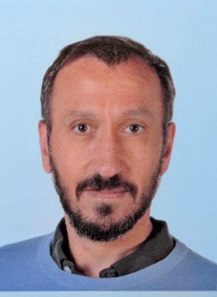
Prof. Gianluca Grenci
Prof. Gianluca Grenci is an Associate Professor in the Department of Industrial Engineering at the University of Padua, joining the faculty in May 2026. Previously, he spent over 13 years at the Mechanobiology Institute (MBI) at the National University of Singapore, where he managed the Micro-fabrication Core Facility and led research in biomedical engineering as a Senior Research Fellow.
His research bridges advanced micro- and nanofabrication with mechanobiology, focusing on microfluidic platforms, organoid microenvironments, and microgravity simulation systems for cell biology and space research.